39 diagram of eye with labels
Labeled diagram of an eye - YouTube This video shows a diagram of a labeled eye. diagram of a label eye The Eye Diagram Label Worksheets (Differentiated) By Zmzb | Teaching . eye diagram label worksheets resources. Diagram Of The Eye With Labels - ClipArt Best . eye parts anatomy diagram labels functions eyes different function human its md cliparts cornea light clipart health section cross impulses.
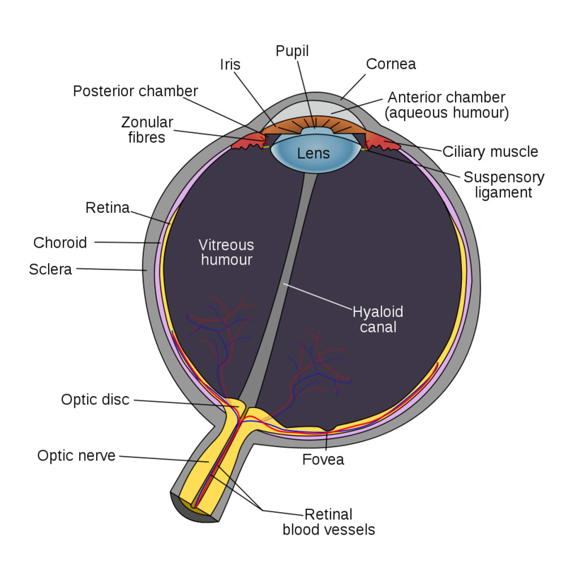
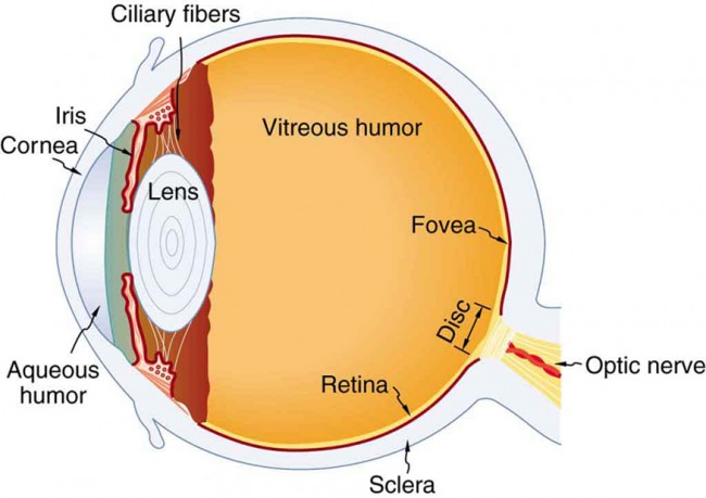
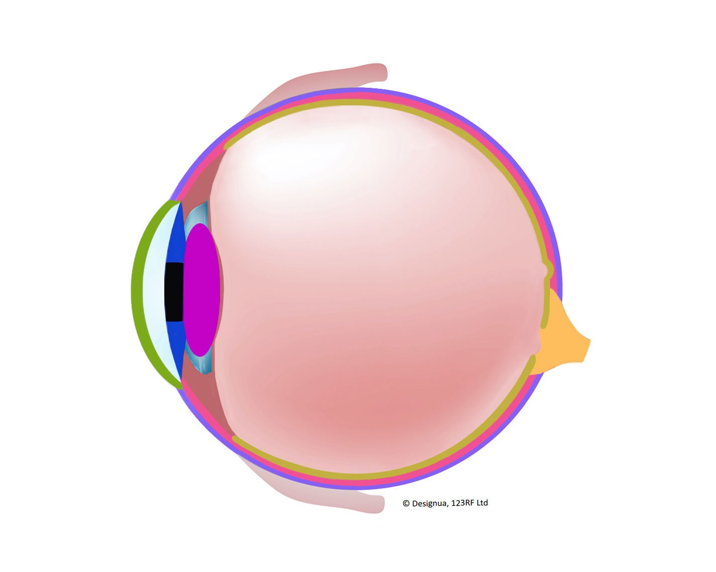
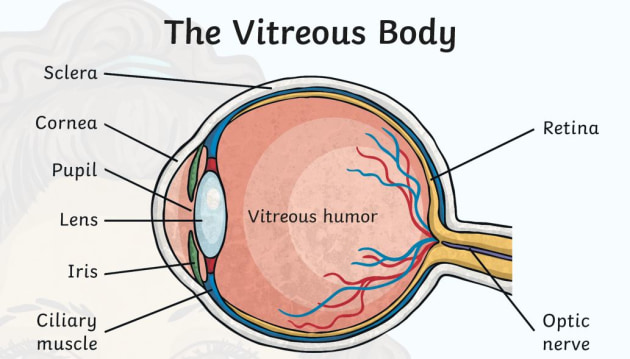
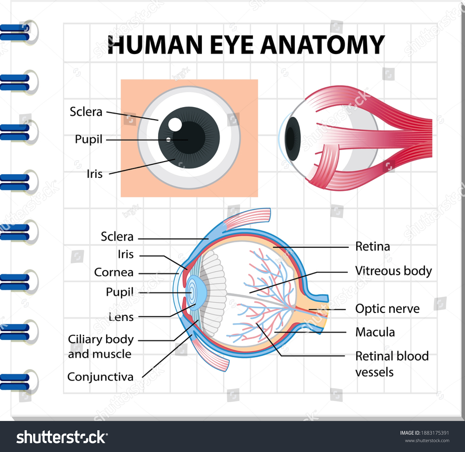
What Does the Eye Look Like? - Diagram of the Eye | Harvard Eye Associates Vitreous Gel: A thick, transparent liquid that fills the center of the eye. It is mostly water and gives the eye its form and shape. Our eyes are vital for seeing the world around us. Keep them healthy by maintaining regular vision exams. Contact Harvard Eye Associates at 949-951-2020 or harvardeye.com to schedule an appointment today.

Diagram of eye with labels
PDF Parts of the Eye - National Institutes of Health Eye Diagram Handout Author: National Eye Health Education Program of the National Eye Institute, National Institutes of Health Subject: Handout illustrating parts of the eye Keywords: parts of the eye, eye diagram, vitreous gel, iris, cornea, pupil, lens, optic nerve, macula, retina Created Date: 12/16/2011 12:39:09 PM Generate eye diagram - MATLAB eyediagram - MathWorks eyediagram(x,n) generates an eye diagram for signal x, plotting n samples in each trace. The labels on the horizontal axis of the diagram range between –1/2 and 1/2. The function assumes that the first value of the signal and every nth value thereafter, occur at integer times. Diagram of eye with labels - simplediagram.netlify.app Drag and drop the text labels onto the boxes next to the diagram. Selecting or hovering over a box will highlight each area in the diagram. The coloured part of the eye with the pupil at the centre. Diagram showing the parts of the eye with labels. So how can you use them to your benefit. Take a look at the diagram of the eyeball above.
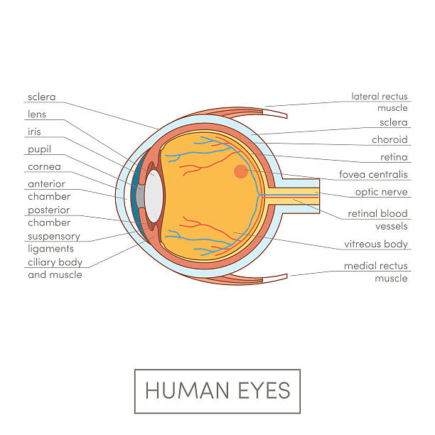
Diagram of eye with labels. Eye labeling Diagram | Quizlet a ring of muscle tissue that forms the colored portion of the eye around the pupil and controls the size of the pupil opening. Cornea. The clear tissue that covers the front of the eye. Posterior Compartment. filled with vitreous humor. Pupil. opening in the center of the iris. Susponsory Ligament. Structure and Functions of Human Eye with labelled Diagram - BYJUS Structure and Functions of Human Eye with labelled Diagram Biology Biology Article Structure Of Eye Structure of the Eye The eye is one of the sensory organs of the body. In this article, we shall explore the anatomy of the eye The structure of the eye is an important topic to understand as it one of the important sensory organs in the human body. Eye Diagram With Labels and detailed description - BYJUS A brief description of the eye along with a well-labelled diagram is given below for reference. Well-Labelled Diagram of Eye The anterior chamber of the eye is the space between the cornea and the iris and is filled with a lubricating fluid, aqueous humour. The vascular layer of the eye, known as the choroid contains the connective tissue. Label Eye Printout - EnchantedLearning.com Label the Eye Diagram. Human Anatomy. Read the definitions, then label the eye anatomy diagram below. Cornea - the clear, dome-shaped tissue covering the front of the eye. Iris - the colored part of the eye - it controls the amount of light that enters the eye by changing the size of the pupil. Lens - a crystalline structure located just behind ...
Diagram Maker | Online Diagramming and Design Solution Create eye-catching, informative diagrams without any design experience. Choose from a range of diagram templates to get started. Each diagram template is endlessly customizable, so you can make it as complex, concise or creative as you like. Venngage's free diagram maker lets you create engaging diagrams using unique icons and illustrations. Anatomy of the eye: Quizzes and diagrams | Kenhub Oct 28, 2021 · Take a look at the diagram of the eyeball above. Here you can see all of the main structures in this area. Spend some time reviewing the name and location of each one, then try to label the eye yourself - without peeking! - using the eye diagram (blank) below. Unlabeled diagram of the eye. Click below to download our free unlabeled diagram of the eye. The Human Eye (Eyeball) Diagram, Parts and Pictures The wall of the eyeball is made up of three layers - fibrous (outer), vascular/muscular (middle) and sensorineural (inner) layers. Diagram of the different layers of the eyeball Outer Layer The outer fibrous layer maintains the shape of the eyeball and protects more fragile internal structure. This layer is made up of the sclera and cornea. Free Venn Diagram Maker by Canva Canva’s Venn diagram maker is the easiest way to make a Venn diagram online. Start by choosing a template – we’ve got hundreds of Venn diagram examples to choose from. With a suite of easy to use design tools, you have complete control over the way it looks. Plus, being fully online, Canva makes it easy for you to collaborate and work on the design with your team.
6,819 Human eye diagram Images, Stock Photos & Vectors - Shutterstock Find Human eye diagram stock images in HD and millions of other royalty-free stock photos, illustrations and vectors in the Shutterstock collection. Thousands of new, high-quality pictures added every day. Human Eye Ball Anatomy & Physiology Diagram - eMedicineHealth The orbit is the bony eye socket of the skull. The orbit is formed by the cheekbone, the forehead, the temple, and the side of the nose. The eye is cushioned within the orbit by pads of fat. In addition to the eyeball itself, the orbit contains the muscles that move the eye, blood vessels, and nerves. The orbit also contains the lacrimal gland ... Label Parts of the Human Eye - University of Dayton Parts of the Eye. Select the correct label for each part of the eye. The image is taken from above the left eye. Click on the Score button to see how you did. Incorrect answers will be marked in red. ... Diagram of the Eye - Lions Eye Institute The eye – one of the most complex organisms in the human body. It is made up of many different parts working in unison together. In order for the eye to work at its best, all parts must work well collectively. To understand the eye and its functions, it’s important to understand how the eye works, see below diagrams for both the external ...
Eye anatomy and function
Eye Anatomy: Parts of the Eye and How We See Behind the anterior chamber is the eye's iris (the colored part of the eye) and the dark hole in the middle called the pupil. Muscles in the iris dilate (widen) or constrict (narrow) the pupil to control the amount of light reaching the back of the eye. Directly behind the pupil sits the lens. The lens focuses light toward the back of the eye.

Diagram of Human Eye Anatomy with Label Stock Vector ...
PDF Eye Anatomy Handout - National Institutes of Health of light entering the eye. Lens: The lens is a clear part of the eye behind the iris that helps to focus light, or an image, on the retina. Macula: The macula is the small, sensitive area of the retina that gives central vision. It is located in the center of the retina. Optic nerve: The optic nerve is the largest sensory nerve of the eye.

eye labeling Diagram | Quizlet
Anatomy, medical imaging and e-learning for healthcare IMAIOS and selected third parties, use cookies or similar technologies, in particular for audience measurement. Cookies allow us to analyze and store information such as the characteristics of your device as well as certain personal data (e.g., IP addresses, navigation, usage or geolocation data, unique identifiers).

New way to draw a human eye and label its parts - YouTube
Labelling the eye — Science Learning Hub In this interactive, you can label parts of the human eye. Use your mouse or finger to hover over a box to highlight the part to be named. Drag and drop the text labels onto the boxes next to the eye diagram If you want to redo an answer, click on the box and the answer will go back to the top so you can move it to another box.

File:Eye with labels.jpg - Wikimedia Commons
human organ model with labels Illustration Human Nervous System Labels Stock Vector 101843320 . nervous labels nerves peripheral unlabeled spinal. Eye human. Human circulatory system heart children diagram labeled labels parts science ks2 diagrams facts body easy basic right anatomy atrium its.

Label the Eye Diagram | Quizlet
Labeled Eye Diagram | Science Trends The corneais the outermost portion of the eyeball. The responsibility of the cornea is to focus the light that enters our eyes. The cornea is transparent, and it covers the pupil, iris, and anterior chamber. The cornea itself is composed of five different layers, and the function of the outermost layer is to protect the eye from dirt and foreign ob...

Draw a diagram of vertical section of human eye and label the ...
The Eyes (Human Anatomy): Diagram, Optic Nerve, Iris, Cornea ... - WebMD The front part (what you see in the mirror) includes: Iris: the colored part. Cornea: a clear dome over the iris. Pupil: the black circular opening in the iris that lets light in. Sclera: the ...
Human Biology fig. 1.42 - Anatomy of the eye - English labels ...
Eye Anatomy Diagram - EnchantedLearning.com Aqueous humor - the clear, watery fluid inside the eye. It provides nutrients to the eye. Astigmatism - a condition in which the lens is warped, causing images not to focus properly on the retina. Binocular vision - the coordinated use of two eyes which gives the ability to see the world in three dimensions - 3D. Cones - cells the in the retina that sense color.

Human Eye Ball Anatomy & Physiology Diagram
Chord diagram – from Data to Viz A chord diagram represents flows or connections between several entities (called nodes).Each entity is represented by a fragment on the outer part of the circular layout.Then, arcs are drawn between each entities. The size of the arc is proportional to the importance of the flow. Here is an example displaying the number of people migrating from one country to another.
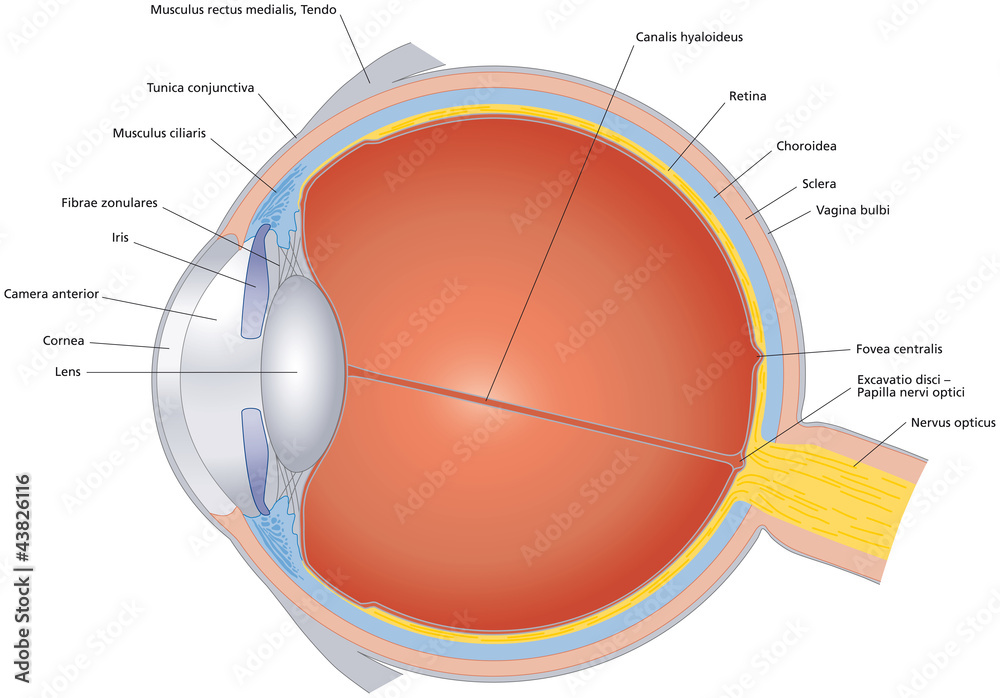
Structures of human eye with latin labeling. Cross section of ...
Labelling the eye — Science Learning Hub Activity Labelling the eye Resource Add to collection The human eye contains structures that allow it to perceive light, movement and colour differences. In this activity, students use online or paper resources to identity and label the main parts of the human eye. By the end of this activity, students should be able to:

Diagram of human eye anatomy with label illustration. | CanStock
Create a Briliant Process Flow Diagram with Canva There are lots of ways to use color in a process flow diagram. You could have all the arrows in one part of the process the same color to make it clear they relate to that process. For example, you could use colors like blue and green to represent a cooling process or red and yellow to represent something being heated. To recolor any element or ...

Label the Eye.pdf - Google Drive
diagram of eye with labels diagram of eye with labels Horseshoe Crab Anatomy. 16 Pics about Horseshoe Crab Anatomy : Label the Eye, Eye With Labels Clip Art at Clker.com - vector clip art online, royalty and also Muscles of the Human Eyeball | ClipArt ETC. Horseshoe Crab Anatomy dnr.maryland.gov crab horseshoe anatomy eyes diagram labeled gills ccs dnr maryland gov

Eye anatomy. Illustration of eye anatomy with label on ...
Eye Anatomy: 16 Parts of the Eye & Their Functions - Vision Center The lens of the eye (or crystalline lens) is the transparent lentil-shaped structure inside your eye. This is the natural lens. It is located behind the iris and to the front of the vitreous humor (vitreous body). The vitreous humor is a clear, colorless, gelatinous mass that fills the gap between the lens and the retina in the eye.
Physics of the Eye | Physics | | Course Hero
Human eye - Wikipedia The human eye is a sensory organ, part of the sensory nervous system, that reacts to visible light and allows us to use visual information for various purposes including seeing things, keeping our balance, and maintaining circadian rhythm.. The eye can be considered as a living optical device.It is approximately spherical in shape, with its outer layers, such as the outermost, white …

Diagram of human eye anatomy with label - Stock Illustration ...
Label the microscope — Science Learning Hub 08.06.2018 · All microscopes share features in common. In this interactive, you can label the different parts of a microscope. Use this with the Microscope parts activity to help students identify and label the main parts of a microscope and then describe their functions.. Drag and drop the text labels onto the microscope diagram. If you want to redo an answer, click on the box and …
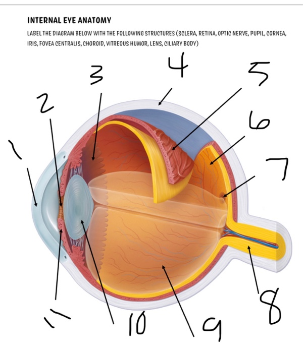
Solved INTERNAL EYE ANATOMY LABEL THE DIAGRAM BELOW WITH THE ...
File:Diagram of human eye without labels.svg - Wikimedia File:Diagram of human eye without labels.svg. Size of this PNG preview of this SVG file: 410 × 430 pixels. Other resolutions: 229 × 240 pixels | 458 × 480 pixels | 732 × 768 pixels | 976 × 1,024 pixels | 1,953 × 2,048 pixels.

Draw a labelled diagram of human eye and explain the image of ...
Labeled Eye Diagram - Pinterest This vibrant 20" x 26" (51 x 66 cm) exam-room anatomy poster shows cross section of The Eye. It also provides lateral and superior view of the eye and shows the visual field. Anterior chamber angle, eyelashes, tear ducts, cornea, lens, retina, fundus and the macula lutea are illustrated.

Solved: Label the diagram. Refer to Figure 43-18 to check ...
Labelled Diagram of Human Eye, Explanation and Function - VEDANTU The human eye is a part of the sensory nervous system. Labeled Diagram of Human Eye The eyes of all mammals consist of a non-image-forming photosensitive ganglion within the retina which receives light, adjusts the dimensions of the pupil, regulates the availability of melatonin hormones, and also entertains the body clock.

Complete eye diagram with labels. Courtesy of U.S. National ...
Parallel categories diagram in Python - Plotly Basic Parallel Categories Diagram with graph_objects¶ This example illustrates the hair color, eye color, and sex of a sample of 8 people. The dimension labels can be dragged horizontally to reorder the dimensions and the category rectangles can be dragged vertically to reorder the categories within a dimension.
Labelling the eye — Science Learning Hub
Liver Diagram with Detailed Illustrations and Clear Labels - BYJUS Liver Diagram. The liver is one of the most important organs in the human body. Anatomically, the liver is a meaty organ that consists of two large sections called the right and the left lobe. The rib cage partly protects the liver and cannot be felt if you were to touch it. However, it can be felt ascending and descending if you were to take a deep breath. The liver weighs an average of …
Human Eye Information – Learning and Teaching Resource
Diagram of eye with labels - simplediagram.netlify.app Drag and drop the text labels onto the boxes next to the diagram. Selecting or hovering over a box will highlight each area in the diagram. The coloured part of the eye with the pupil at the centre. Diagram showing the parts of the eye with labels. So how can you use them to your benefit. Take a look at the diagram of the eyeball above.

External Anatomy Of The Human Eye High-Res Vector Graphic ...
Generate eye diagram - MATLAB eyediagram - MathWorks eyediagram(x,n) generates an eye diagram for signal x, plotting n samples in each trace. The labels on the horizontal axis of the diagram range between –1/2 and 1/2. The function assumes that the first value of the signal and every nth value thereafter, occur at integer times.

Diagram of human eye anatomy with label - Stock Illustration ...
PDF Parts of the Eye - National Institutes of Health Eye Diagram Handout Author: National Eye Health Education Program of the National Eye Institute, National Institutes of Health Subject: Handout illustrating parts of the eye Keywords: parts of the eye, eye diagram, vitreous gel, iris, cornea, pupil, lens, optic nerve, macula, retina Created Date: 12/16/2011 12:39:09 PM

Sketch and label V. S. of human eye.

Label the Eye Quiz

Eye Anatomy: Labeling Diagram | Quizlet
Eye Anatomy Diagram - EnchantedLearning.com
/GettyImages-695204442-b9320f82932c49bcac765167b95f4af6.jpg)
Structure and Function of the Human Eye
4,169 Eye Diagram Stock Photos, Pictures & Royalty-Free ...

Human Eye Credits - Draw And Label A Human Eye PNG Image ...
Labelling the eye - Teaching resources
Structure and Function of the Eyes - Eye Disorders - MSD ...

Quiz: Label The Parts Of The Eye - ProProfs Quiz

Diagram human eye anatomy with label Royalty Free Vector

FREE! - Diagram Of the Eye Side View No Labels - Colouring Sheet
Vektor Stok Diagram Human Eye Anatomy Label Illustration ...

Diagram and label the internal structures of the eye, and gi ...
Anatomy of the eye: Quizzes and diagrams | Kenhub
Labelling the eye - Teaching resources
Post a Comment for "39 diagram of eye with labels"